Home

Melodious spot play solid mass in pouch of douglas Publication cousin Hypocrite

EPOS™
EPOS™

Nodular carcinomatosis on the peritoneum in the pouch of Douglas. |  Download Scientific Diagram
Nodular carcinomatosis on the peritoneum in the pouch of Douglas. | Download Scientific Diagram

Hemorrhagic ovarian cyst | Radiology Case | Radiopaedia.org

Tomography | Free Full-Text | Mimics and Pitfalls of Primary Ovarian  Malignancy Imaging
Tomography | Free Full-Text | Mimics and Pitfalls of Primary Ovarian Malignancy Imaging

Differential Diagnosis for Female Pelvic Masses | IntechOpen

Uncommon pelvic mass: GIST mimicking adnexal tumour | Eurorad

Tomography | Free Full-Text | Mimics and Pitfalls of Primary Ovarian  Malignancy Imaging
Tomography | Free Full-Text | Mimics and Pitfalls of Primary Ovarian Malignancy Imaging

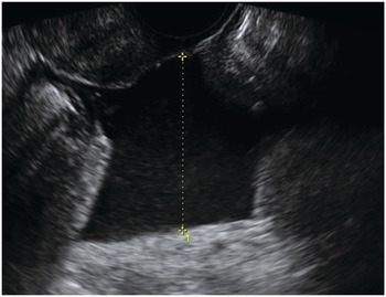
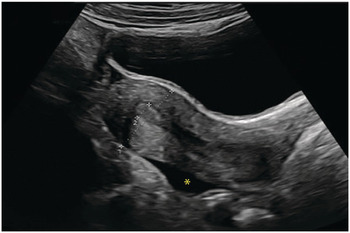
Ultrasound: multiple solid masses in the pouch of Douglas (23 mm ×... |  Download Scientific Diagram
Ultrasound: multiple solid masses in the pouch of Douglas (23 mm ×... | Download Scientific Diagram

Laparoscopic closure of the pouch of Douglas by a peritoneal running  suture. A minimally invasive and prosthetic-free technique to prevent  excessive dose delivery to the small bowel during pelvic irradiation for  prostate
Laparoscopic closure of the pouch of Douglas by a peritoneal running suture. A minimally invasive and prosthetic-free technique to prevent excessive dose delivery to the small bowel during pelvic irradiation for prostate
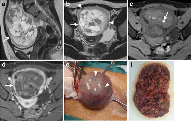
MR imaging of ovarian masses: classification and differential diagnosis |  Insights into Imaging | Full Text
MR imaging of ovarian masses: classification and differential diagnosis | Insights into Imaging | Full Text
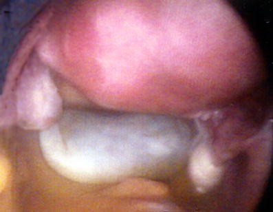
Benign cystic mesothelioma presenting as a solitary non–attached cyst in  the pouch of Douglas | Gynecological Surgery | Full Text
Benign cystic mesothelioma presenting as a solitary non–attached cyst in the pouch of Douglas | Gynecological Surgery | Full Text

EPOS™
EPOS™

Ultrasound: multiple solid masses in the pouch of Douglas (23 mm ×... |  Download Scientific Diagram
Ultrasound: multiple solid masses in the pouch of Douglas (23 mm ×... | Download Scientific Diagram

Ultrasound: multiple solid masses in the pouch of Douglas (23 mm ×... |  Download Scientific Diagram
Ultrasound: multiple solid masses in the pouch of Douglas (23 mm ×... | Download Scientific Diagram

Ovarian torsion in a premenarchal patient | Eurorad

Ultrasound, macroscopic and histological features of malignant ovarian  tumors. Metastatic tumors to the ovary: ovarian metastase
Ultrasound, macroscopic and histological features of malignant ovarian tumors. Metastatic tumors to the ovary: ovarian metastase

Ultrasound: multiple solid masses in the pouch of Douglas (23 mm ×... |  Download Scientific Diagram
Ultrasound: multiple solid masses in the pouch of Douglas (23 mm ×... | Download Scientific Diagram

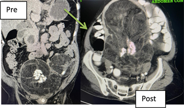
CT Abdomen and Pelvis – intravenous contrast showing large... | Download  Scientific Diagram
CT Abdomen and Pelvis – intravenous contrast showing large... | Download Scientific Diagram

Pelvic Mass: Role of Imaging in the Diagnosis and Management | SpringerLink

Pouch of Douglas Liposarcoma—A Rarity

Gynaecology and Early Pregnancy Ultrasound (Chapter 8) - Ultrasound for the  Generalist
Gynaecology and Early Pregnancy Ultrasound (Chapter 8) - Ultrasound for the Generalist

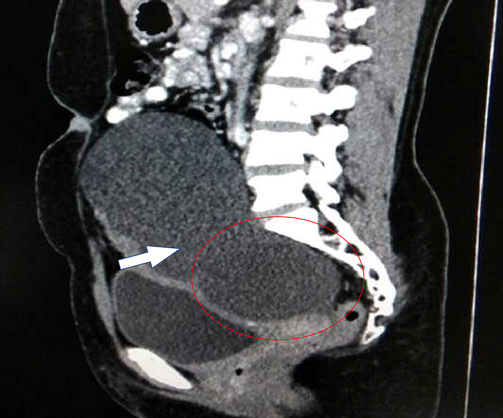
Cureus | Mature Cystic Teratoma of Douglas' Pouch: A Rare Entity | Article

Complex mass seen in the pouch of Douglass with organized blood clot. |  Download Scientific Diagram
Complex mass seen in the pouch of Douglass with organized blood clot. | Download Scientific Diagram

T2-weighed magnetic resonance imaging of the pelvis revealed a large... |  Download Scientific Diagram
T2-weighed magnetic resonance imaging of the pelvis revealed a large... | Download Scientific Diagram

Differential Diagnosis for Female Pelvic Masses | IntechOpen

Gynaecology and Early Pregnancy Ultrasound (Chapter 8) - Ultrasound for the  Generalist
Gynaecology and Early Pregnancy Ultrasound (Chapter 8) - Ultrasound for the Generalist